View all Close all
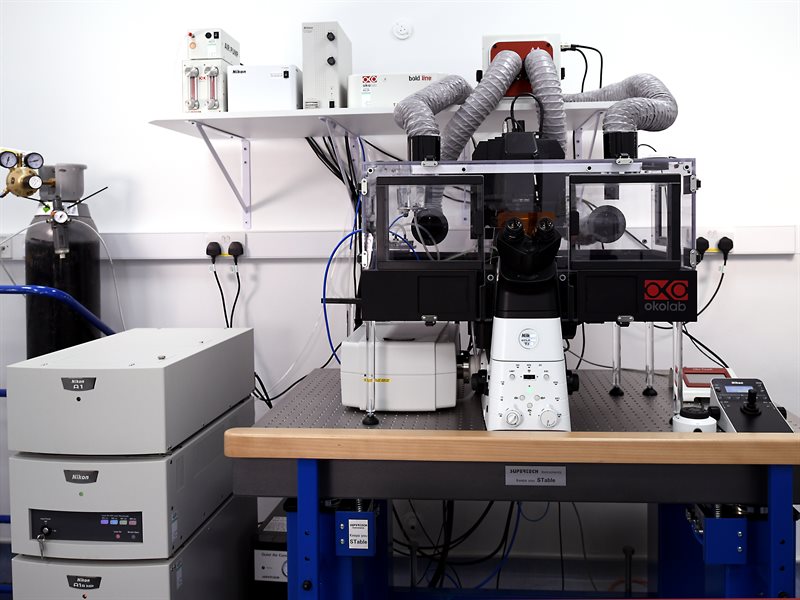
The Nikon A1R confocal microscope at the facility is an automated inverted system featuring a dual scanhead design, enabling both Galvano mode for high-resolution imaging and Resonant mode for high-speed acquisition. Its highly sensitive GaAsp detectors ensure clear, high-resolution images, even from faint signals.
Equipped with an Okolab environmental chamber and stage-top incubator, the microscope allows precise control of temperature and gases, ideal for time-lapse imaging of live samples. It also includes various sample holders for glass slides, Ibidi plates, Petri dishes and microwell plates. We recommend using a cover slip with thickness of 0.17mm when using this equipment.
The Nikon A1R includes the following objectives:
- 10x CFI Plan Fluor DLL (0.3 NA, Ph1, WD: 16 mm)
- 20x CFI Plan Apochromat VC (0.75 NA, WD: 1 mm)
- 40x CFI S Plan Fluor ELWD (WD: 2.8-3.6 mm, correction collar: 0-2.0 mm)
- 60x Oil CFI Apochromat Lambda S (1.42 NA, WD: 0.15 mm, chromatic correction: 405-656 nm)
- 100x Oil CFI Plan Apochromat Lambda D (1.45 NA, WD: 0.13 mm, chromatic correction: 435-850 nm)
- 60x Water CFI Apochromat IR (1.27 NA, WD: 0.17 mm, chromatic correction: 435-1064 nm)
Image acquisition and analysis are streamlined with Nikon NIS-Elements C software, enabling users to go from data collection to publication-ready figures in one session.
For advanced image processing, the system includes a deconvolution module as well as the NIS.AI suites, which features Enhance.ai, Segment.ai, Clarify.ai and Convert.ai.
Book Nikon A1R
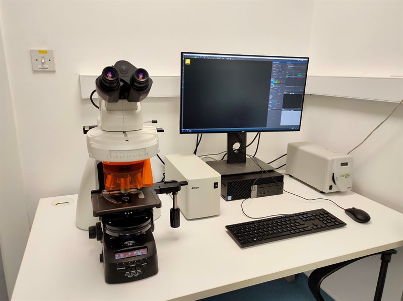
The Nikon NiE is a modern upright research widefield microscope equipped with a Nikon DS-Fi3 camera for both color and emulated monochrome imaging. This allows for high-resolution brightfield and fluorescent imaging. The CoolLED pE300, a broad-spectrum LED fluorescent light source, provides excellent illumination for a wide range of applications.
The NiE includes the follwoing objectives;
- 10x Plan Fluor DLL (NA: 0.3, Ph1, WD: 16 mm)
- 20x Plan Fluor (NA:0.66, DIC, WD: 2.1 mm)
- 40x CFI Plan Fluor (NA: 0.75, WD: 0.66 mm)
- 60x oil Plan Fluor Lambda (NA: 1.40, DIC, WD: 1.3)
Controlled by Nikon NIS-Elements software, the NiE is user-friendly and ideal for viewing slides. It features five fluorescent filters—DAPI, FITC, TRITC, CY3, and CY5—providing versatility for automatic fluorescence image capture and analysis.
Book Nikon NiE
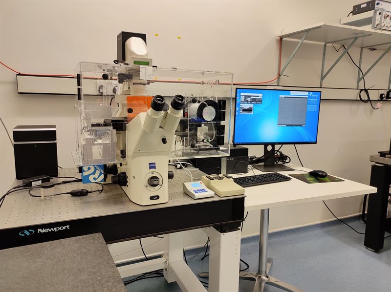
The Axiovert 200M chassis has been fitted with a CoolLED PE300-Ultra LED light source for fast and reliable fluorescent imaging. The Retiga R6 EM-CCD camera provides high speed and high sensitivity, and the automated stage and environmental control chamber facilitates reliable live cell imaging.
Controlled via MicroManager software this platform is very flexible with all the advantages of a modern inverted widefield system.
Book Axiovert 200M
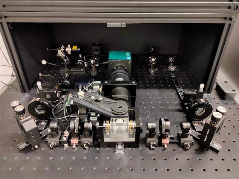
The T-SPIM is a custom-built light sheet microscope designed with dual-sided illumination, ensuring even excitation of large samples such as zebrafish embryos and spheroids. Imaging occurs in a sealed chamber where the sample is immersed in a selected medium, such as PBS or cell culture medium.
T-SPIM (Selective Plane Illumination Microscopy) combines the optical sectioning capabilities of a confocal microscope with the speed and sensitivity of widefield imaging. By exciting the sample with a sheet of laser light, T-SPIM selectively illuminates only the imaging plane, minimizing photobleaching and reducing off-plane excitation, which can impair image resolution in conventional widefield microscopy.
The system supports 10x, 20x, or 40x detection objectives, depending on your sample. Please discuss your imaging needs with IRF staff in advance of your session.
Book T-SPIM Light sheet
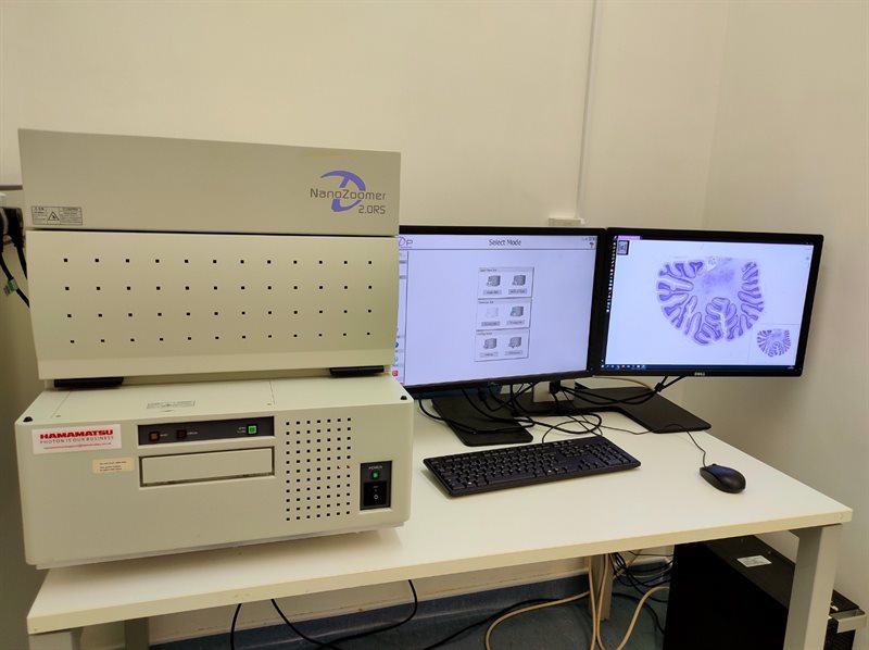
The NanoZoomer 2.0-RS slide scanner from Hamamatsu allows the scanning of up to six standard (76mm x 26mm) or two double-size (76mm x 52mm) slides in a variety of modes: manual, semi-automatic, and automatic.
Scanning can be done at 20x (0.46 µm/pixel) and 40x (0.23 µm/pixel) magnification, with scan times as fast as 1 minute 40 seconds for a 20mm by 20mm area at 20x magnification, with 40x magnification increasing the scan time proportionally.
This system allows the generation of high-resolution images of the entire slide, allowing image analysis to be done when required by the user, without the risk of missing information during image acquisition, since the entire slide is recorded.
The free NDP.view2 software allows viewing, annotation, and export of scans. NDP.Serve allows the hosting of slides online for viewing via a web browser.
Book Nanozoomer
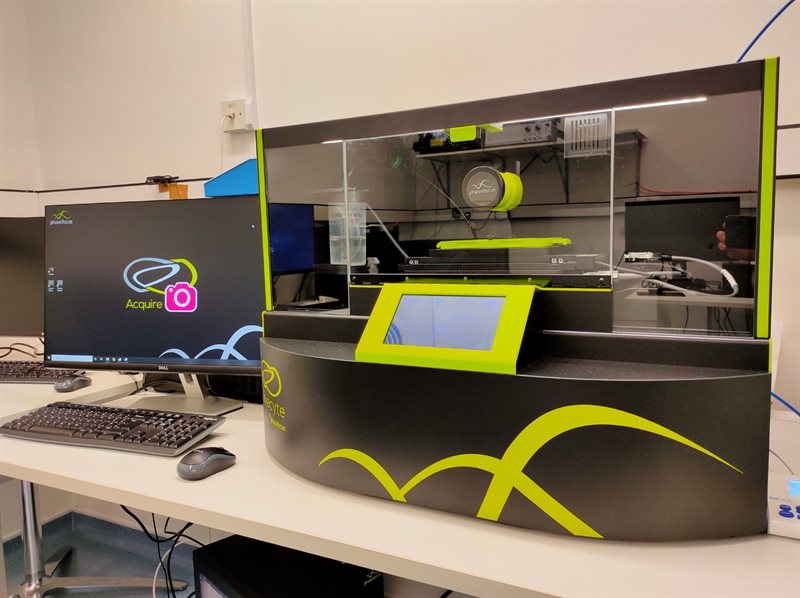
The LiveCyte2 utilises Quantitative Phase Imaging (QPI) to generate label-free high contrast images for automatic quantification of live cells. The proprietary QPI technology allows the gentle imaging of even sensitive cell lines that would normally be prohibitive of live-labelled imaging.
The user-friendly software allows real-time analysis of live time-series experiments, meaning users can set up assays and collect fully processed figure-ready data at the end of the run, along with viable cells that can be passaged or processed for further experimentation.
Phase Focus provide a suite of analysis assays, for tracking a range of metrics in a variety of combinations, with applications in Cell Proliferation and Growth, Wound Healing, Cell Motility, Oncology, Toxicology, Angiogenesis and Stem Cell differentiation.
Book LiveCyte
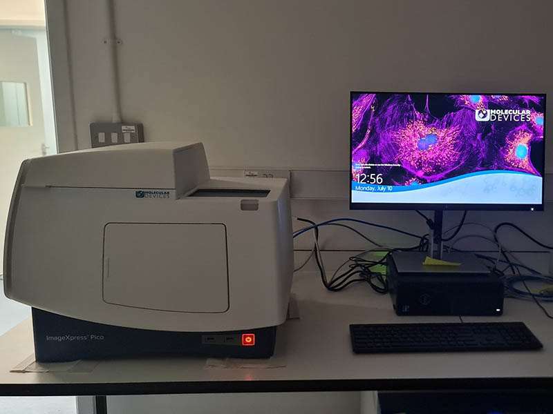
The ImageXpress Pico is a high-throughput widefield microscope for image acquisition in microwell plates and slides. It offers environmental control, including temperature and humidity, to maintain optimal conditions for live cell imaging experiments.
The ImageXpress Pico is equipped with a 1.3-megapixel sCMOS camera that can capture high-resolution images with low noise and high sensitivity. The system supports a wide range of fluorescence dyes and can be used for various fluorescence imaging assays. The system combines advanced optics, live cell imaging capabilities, and user-friendly software to provide high-quality images and efficient data analysis.
The software includes a range of analysis tools for quantifying cell morphology, measuring fluorescence intensity, and performing advanced image processing tasks such as mitochondria assay, angiogenesis, cell and nuclei count etc. It also allows the creation of customisable analysis workflows to automate image analysis and data extraction.
Book ImageXpress Pico
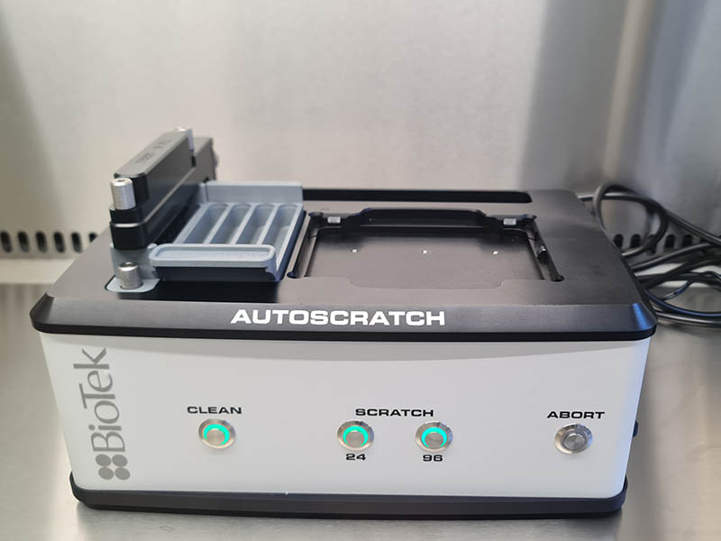
The BioTek autoscratch is an automated wound making tool that create a uniform monolayer of wound in 24 and 96 wells microplate using a single click. The autoscratch is permanently housed inside a class II biological safety cabinet. The device ensures reproducible wound size for wound healing assay.
Book AutoScratch