Image analysis is essential to generate high quality quantifiable data from microscopy images – it is as vital a part of the process as good sample preparation and imaging technique.
The Image Resource Facility provides users with access to analysis software for the processing of images for quantification purposes. Advice about how best to tackle your image analysis needs is available by contacting irf@sgul.ac.uk.
FIJI
For routine visualisation and analysis of microscopy images we recommend the use of FIJI, a free open-source image analysis platform that will open most file types and run on any system. Here are some ImageJ tutorials to get you started with FIJI.
NIS-Elements
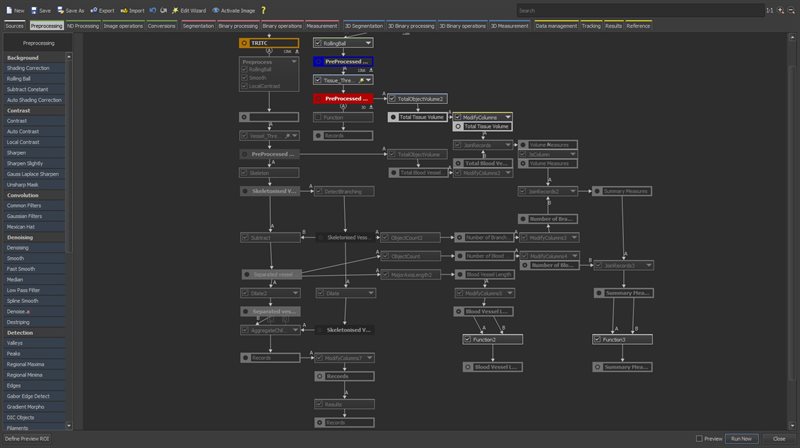
NIS-Elements from Nikon allows for powerful image processing and analysis to be carried out on larger confocal datasets, including deconvolution and denoising. The included JOBS and General Analysis package of NIS-Elements allows the user to set up detailed granular image analysis that can be run in large batches on any number of files, allowing for powerful automation of otherwise time-consuming image processing and analysis.
PhaseFocus Analyse
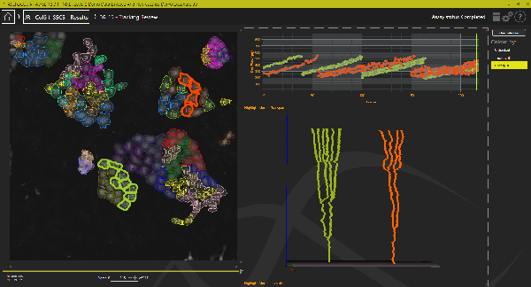
The Analyse software from PhaseFocus is used to process Pytography images generated by the LiveCyte system – once configured, custom analyses can be run in real-time with image acquisition to provide users with their data at the time samples are collected after an imaging run.
Custom Dashboards can be generated to present specific metrics together for any given dataset.