Published: 06 April 2023
Adding a third routine scan at the end of pregnancy can slash the number of unexpected breech births by 70% and the risks of the baby being born with severe health complications, according to research led by doctors at St George’s, University of London, and published in PLOS Medicine.
Call for change to guidelines
The researchers now hope that these findings will lead to a change in NICE guidelines so all pregnant women are offered a scan in their third trimester to improve maternity care.
Currently, pregnant women have routine scans at 12 and 20 weeks only, with no routine scan offered in the third (final) trimester. It is only if they are flagged at risk of a complicated pregnancy (such as having diabetes or high blood pressure) at one of these two scans or antenatal visits that they are referred for further observation.
However, around 4% of babies are unexpectedly in a breech position at the end of pregnancy. This is where the baby is positioned feet or bottom first which puts them at increased risk of being admitted to the neonatal unit, brain injury due to a lack of oxygen, or even death.
“It’s vital we know how the baby is lying towards the end of pregnancy as we want to avoid a breech birth if at all possible. The two routine scans are far too early to tell us how the baby will be positioned at the time of labour and that’s why a third scan at 36-37 weeks could be a game-changer to pregnancy and birth care.”
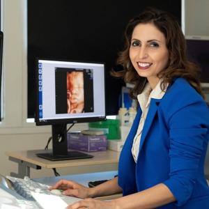
- Professor Asma Khalil, Professor of Obstetrics and Maternal Fetal Medicine at St George’s, University of London, who led the study -
Doctors compared the rate of unexpected breech births and the health of the newborn baby after different third trimester scan policies were introduced at St George’s University Hospital NHS Foundation Trust (SGUH) and Norfolk and Norwich University Hospital NHS Foundation Trust (NNUH).
A total of 24,128 pregnant women were recruited at SGUH, of which 16,777 received only the usual antenatal ultrasound scans and 7,351 had an extra ultrasound scan by a sonographer at 36 weeks. At NNUH, 9,694 women were recruited of which 5,119 women received standard ultrasound scans and 4,575 were given a ‘point-of-care’ ultrasound scan at 36 weeks using a cheap hand-held, portable device that brings the scan up on a phone or tablet.
Drop in complications to mother and baby
Both types of third trimester ultrasound scan dramatically reduced the rate of unexpected breech births – 71% lower with the standard type of ultrasound at SGUH and 69% lower with the hand-held portable device at NNUH.
The babies of women who had the third ultrasound were 16% less likely to be admitted to the neonatal unit for closer monitoring, and were 40-77% less likely to have a low ‘Apgar score’ at 5 minutes after being born. The Apgar score assesses appearance and skin colour, heart rate, reflexes, muscle function, ability to breathe, and is an assessment of the baby’s wellbeing soon after birth. The mothers were also less likely to need an emergency caesarean.
Solution to help improve maternity care
Professor Asma Khalil continues: “For the first time we’ve shown that just one extra scan could save mothers-to-be from trauma, an emergency C-section, and their babies from having severe health complications which could otherwise have been prevented."
“Our research comes at a time when there’s a spotlight on the safety of maternity services and provides the NHS with a clear solution to help enable maternity units better prepare for safer healthier births.”
- Professor Asma Khalil -
Professor Khalil now hopes that more midwives can be trained to use the hand-held ultrasound device so pregnant women can have easy access to a third trimester scan either in the comfort of their own home or at hospital.
Woman-centred care
Fran Harlow, Consultant Obstetrician for the Norfolk and Norwich University Hospital and Maternal Medicine Lead for the Norwich Regional Maternal Medicine Centre, said: “We are delighted that this study has shown that the benefits to the mother and baby are equivalent to a formal scan performed by an ultra-sonographer. This keeps care in the community and woman centred. We hope that the data from NNUH and St George’s provides the stimulus for a national policy of third trimester scanning. In addition, this study has demonstrated that use of a hand-held ultrasound device by a midwife is an innovative, progressive, and now proven, way of achieving this.”
Find out more about our research at the Institute for Molecular and Clinical Sciences